


Onychomycosis is a type of fungal lesion, which affects the exclusive surface of the nail.To prevent the spread of the infection in time, you need to know what looks like a nail fungus, as the therapeutic effect will be achieved faster if the treatment begins in the early stages of the infection.Often, the disease occurs in older people due to poor immunity, but the risk of developing each person's illness.No classification of one fungal nail damage;In medical practice, it is common to distinguish it from the localization and depth they penetrate.The infection is also collected depending on the type of pathogen.
The type of pathogen and the main sign
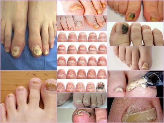
Symptoms of nail fungus in the early stages are easily determined by the condition of the nail.This sign is the most informative, as onychomycosis always manifests itself in the form of changes in surface color, bed deformation, exfoliation and any external changes.The latter is expressed by roughness, grooves and cracks, as well as points that are striked and breached nails.
The main signs of healthy nails are pink and transparency.Onychomycosis at any stage is characterized by nail turmoil and color change to yellow, brown, or green green.In advanced stages, the surface can gain black shade against the background of almost complete destruction.
The signs of external infection with fungus depend on the type of pathogen that is infected.In medical practice, the wounds may be distinguished:
- Candida infection with fungus, which is expressed by nail release directly at the base of the box.Candidiasis of rollers around the nails will be a feature of candidiasis.This version of the complications can have bacterial sources in the form of streptococci or staphylococci, or expressed in the form of a medium plaque or psoriasis;
- Dermatophytes type trichophyton rubrum.In this case, the penetration of the infection occurs directly from the edge of the nail.The first symptom of such pathogens is the appearance of yellowish place on the surface.In the field of neoplasms, the nails collapsed, and the place itself tends to increase.The general place of localization of neoplasms is along the plate, parallel to the nail roller, in this case, the infection is called distal-lateral.There is another form of defeat by this pathogen - distal, where the place appears in the part released from the hole, especially in the middle of the free advantage;
IMPORTANT!
This type of fungus is most common in the toes, gradually making the nails a loose yellow mass.In the absence of proper treatment, the disease flows into hyperkeratosis.The nail plate is completely destroyed due to the spread of the infection around the perimeter.
- Dermatophytes like trichophyton mentagrophytes.Onychomycosis with such filming is also most often based on thumb nails, less often than small fingers.This type of fungal infection requires therapy not only from the nails, but also the feet due to the rapid spread of pathogens.A symptom disease can be like leikonichia, a common disease in medical practice.The main signs are white spots that appear on various parts of the nail, neoplasms are distinguished by non -standard and various sizes.It is easy to distinguish fungus from leanonichia - in the second case, the spots -spots are air collection, which is not observed with fungal damage;
- Mold fungus.This choice of damage is found to be less frequent than candidal or dermatophytic forms.The main sign of such infections - the surface of the plate obtains dark shade, almost black.Not the whole finger can be completely infected, but only part of the nail plate.The first sign of nail fungus on this type of foot is a sharp color change.Onychomycosis can develop in the form of black or dark longitudinal strips in color against the background of the pink nail.
Diagnostics by type of change
It is not difficult to see the nail fungus on the feet even at the main stage of the infection, as this type of infection is actively showing itself from the first day of the wound.From regular transparent nails pale pink in patients, significant surface deformation and general changes in observed conditions.The affected area has a dull yellowish yellow color, which appears primarily in the thumb.The type of fungus and the level of damage are determining the factor.
In the first stage, fungal damage to the feet in the leg is a small foci appearance.The thickening of the plate and keratinization of the bed under the nail will be a feature.This level is accompanied by phenomena such as partial detachment and nail plate release, which acts as a source of infection for healthy people.
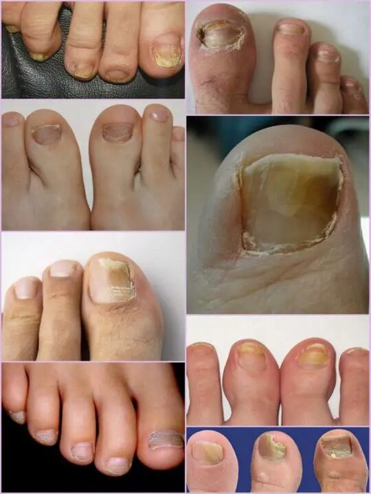
Although the thickening of the plate is active, continuous grinding can be observed, regardless of current factors.The features of each level and symptoms of the fungus in the foot depend directly on the type of pathogen.
Depending on the changes that occur with the nail plate, three options for onychomycosis are distinguished:
- Hypertrophic;
- Normotrophic;
- Atopic.
In the first case, there is a sharp change under the shade of the nail plate, its destruction along the edges, and the deformation of the plate surface.Nails thicken so much that cause discomfort and painful sensations while walking.Normotrophic mycosis of the nails in the foot is distinguished by the presence of a healthy shine, but the plate itself acquires some donkeys, the spots are formed on it.With a type of atropic damage, the focal form of brown and gray on the nail surface.Thanks to them, it is possible to accurately determine the location of the pathogen.
IMPORTANT!
The type of atopic or onycholytic nail fungus is characterized by thinning of the nail plate, and not its growth, as in other cases.The areas where pathogens are localized to escape.Lack of proper treatment leads to advanced stages - complete rejection of the nail plate.
Locations classification
Another sign of which you can separate fungal damage to the toenail depending on the foci location on the nail plate.This also includes pathogenic depth, which in turn allows you to determine the duration of future therapy and speedy recovery opportunities.
Funny fungal disease at localization locations is classified into the following groups:
- Onychomycosis is a type of white surface - the appearance of many white spots on the surface of the nail plate.Fungal infections lead to skin detachment in the appearance of the spots -a spots where there is an active scale release.Advanced levels lead to complete destruction and rejection of plate;
- Distal - develops on the nail -free edge.Color changes were observed first in the small area of the plate, after which there was an active expansion of its boundaries.These lesions are characterized by yellowish or brown gray, as well as uneven surfaces and gradually exfoliating;
- Lateral onychomycosis has a stage of development similar to distal, but it is exclusively localized on the side of the nail plate;
- Total infection - complete infection of the entire surface;
- Proximal onychomycosis.The disease begins its activity from the cuticle, which is inflamed, so the fungus quickly affects the nails, and the process itself begins with the appearance of a small white place near the periolous roller area.
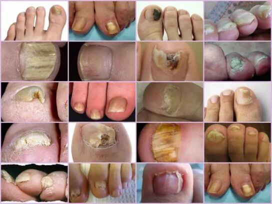
The most common form of nail mycosis on the feet is side and distal, which is usually caused by dermatophytes.Forms of damage such as proximal and white can act as secondary diseases that accompany the immune system disease, for example, Vich.The amount of damage to the nails with fungus should be considered as the advanced stage of any fungus under the nails.
Features -The fungus is under a microscope
Although there are some impressive signs of onychomycosis, other diseases related to skin problems and non -contagious are accepted for fungal damage to the toenails.You can determine the exact diagnosis and type of pathogen only in the laboratory under the microscope, where, in the hospital, the binding of biological materials from the affected area is performed.
The resulting biomaterials are pre -places in the alkaline environment, after which a variety of improvements are made.Studying nail fungus under a microscope allows you to see active pathogens, an external form that determines its type, distribution scale and degree of damage.In the nutrient medium, you can predict the growth of colonies, which allows not only to make the right diagnosis, but also to determine the limitations of the infection.
IMPORTANT!
Since it is possible to determine the presence of onychomycosis only in the laboratory, you cannot postpone a visit to your doctor with little suspicion.Nail fungus develops rapidly, and losing time increases therapy.
What is a worrying signal?
Nail fungus on the feet is indicated by certain symptoms that are partly similar to some skin diseases.The following signs are more likely to show onychomycotic wounds that require medical intervention:
- The appearance of the spots -yellow spots on the plate, its deformation, structural changes, which are not previously complied with;
- Dark plate, loss of transparency, some pictures of nails on the feet show a shade close to black, which is a feature of pathogenic mold;
- Thickening and keratinization of the nail plate, or vice versa is too sharp;
- Rejection of nails from bed, scale detachment and powder;
- Swelling of the roller, hanging it on a plate, fluid release;
- Nails affected by the fungus appear to be inflamed, regardless of the type of pathogen.
All of these symptoms show a high risk of infection.Some external symptoms change the structure of the nail plate can be the result of other diseases.With increased disadvantages, the amount of calcium and iron in the body needs to be increased, increased tuberosity means any body infection, with only white strips and points, copper or zinc deficiency is possible.Although in the photo, the nail fungus looks like a focal damage to the plate with more color changes in yellow, this does not always indicate infection.The yellowness can present a variety of problems with the stomach and liver.
It is not difficult to recognize onychomycosis on the fingers with external signs, despite the extensive classification of the disease.But you can determine the exact type of pathogen and the degree of damage can only be in the laboratory.